what does homogeneous bone marrow signal mean
Chemotherapy and radiation therapy change the marrow in predictable ways and may affect its appearance on MR.
You could call the doctor and ask. Specific, may be present in a number of different conditions or necrosis ( tissue death ) specific may. If you have radiating leg, you should see a spine surgeon for direction on your activity and treatment plan. The corresponding T2w FSE image does not appear particularly abnormal. Overall, 10% of patients with abnormal marrow on MRI were diagnosed with a malignancy. T1w axial image in a normal 26 month-old boy. Bone marrow involvement upstages the patient and may have prognostic and therapeutic implications. ; t exceed 20 % study concluded with the admonition that abnormal bone both red bone marrow ( blood ) > Heterogeneity of the bone marrow activity can be found at the centre of bones! On the other hand, a heterogeneous bone marrow signal can indicate an underlying health condition. 2004; 183:645-653, 28 Kaplan KR, Mitchell DG, Steiner RM, Murphy S, Vinitsi S, Rao VM, Burk L, Rifkin MD: Polycythemia vera and myelofibrosis: Correlation of MR imaging, clinical, and laboratory findings. The most common pathologic infiltration of marrow is metastases from solid organ tumors, but since metastatic disease is much more often multifocal than diffuse in its imaging pattern in the spine, it will only be briefly discussed in this article. It is interesting that some authors emphasize the STIR and post-contrast differences in appearance between hyperplastic red marrow and malignant infiltration while others do not.
Figure 3: The T2-weighted fast spin echo sequence is relatively insensitive to the abnormal marrow, and is largely unremarkable. Perhaps someone else knows. Bone marrow signal of the clivus changes predictably with age and is well assessed with midline T1 non-fat-saturated, non-contrast images. Mri showed bone marrow edema in thoracic spine ?????
Cohort study of patients over age 18 undergoing MRI between what does homogeneous bone marrow signal mean 2005 and October,! Bone marrow cancer is a broad category that includes types such as multiple myeloma. This pattern may persist for many years (10a,11a). Your email address will not be published. - T1 v T2 images - Radiology Masterclass < /a > What does homogeneous signal intensity, intermediate! Red marrow predominates along the periphery. Increased T2-weighted signal from the subchondral bone marrow is a frequent finding in acute traumatic osteochondral injury [86] as well as in the setting of chronic osteochondral injury, or osteoarthritis [87-89]. In adults with normal marrow, the marrow should have higher T1-weighted signal than muscle. For example, a dermoid cyst has heterogeneous attenuation on CT. Radiology.1990; 175:213-218, 31 McKinstry CS, Steiner RE, Young AT, Jones L, Swirsky D, Aber V: Bone marrow in leukemia and aplastic anemia: MR imaging before, during and after treatment. 15, Issue 2, Pages 175-198, 9 Mirowitz S, Apicella P, Reinus WR, Hammerman AM: MR imaging of bone marrow lesions: Relative conspicuousness on T1-weighted, fat-suppressed T2-weighted, and STIR images. Focal areas of red marrow may be a challenge to disclose its nature in some clinical scenarios and mandates making use of different MR pulse sequences to disclose its nature. Pattern 1 begins in younger patients where central fat is visible along the basivertebral veins. reactive marrow: Reactive bone marrow A descriptor for a polyclonal BM response to a local or systemic 'insult', often inflammatory, which may be confined to one cell line, as in reactive granulocytosis, reactive mast cell hyperplasia, reactive thrombocytosis. There is inhomogeneity of the marrow signal throughout the lumbar spine. I had anterior discectomy and fusion at c7 to t 1 because of cervical radiculopathy from a bone spur How common is it for the bone spur to come back and larger after fusion. The use of MR has impacted the staging and follow-up of patients with myeloma and has led to a modification of the classic Durie and Salmon staging system. Addressing bone marrow signal pattern is an integral part of the spinal magnetic resonance (MR) imaging evaluation. Appearance of Normal Marrow in the Spine on MRI. Tumor heterogeneity is one of the hallmarks of malignancy. The T12 and L1 bodies are probably diffusely abnormal for age. This process is referred to as reconversion or sometimes myeloid hyperplasia. What does heterogeneous signal mean on MRI? A T1w sagittal image demonstrates heterogeneous marrow that is generally reduced in signal. Radiology 2004; 231(1):11-23. Maintained height and signal intensity of the other examined discs, with no disc bulges or herniations. However some focal lesions in myeloma do not change significantly in appearance for up to 5 years. Addressing bone marrow signal pattern is an integral part of the spinal magnetic resonance (MR) imaging evaluation. How can a map enhance your understanding? AJR Am J Roentgenol. When an mri shows abnormal bone marrow signals in the hip, there could be a number of causes. A T1w axial image (14c) demonstrates that marrow signal in sacrum and iliac bones is lower than that of adjacent muscle. Become a Gold Supporter and see no third-party ads. Other causes include: hyperplastic anemias such as sickle cell disease, thalassemia and spherocytosis(14a,14b,14c,15a); administration of erythrocyte or granulocyte stimulating agents for therapy and polycythemia vera (16a). Bone marrow edema is an area of increased fluid inside the bone. What does please be guided accordingly phrase means? ; They may be present in a wide range of conditions. These observations plus the fact that reconversion tends to be focal has contributed to some MR descriptions of aplastic anemia as heterogeneous. It is important to note that the introduction of each of these special techniques has met with some success and some disappointment.
Edema probably from bone infarcts is highlighted by arrows in 14a and 14b. The variegated pattern has been described as if cracked pepper were sprinkled onto the marrow on T1-weighted images1,8. The spine is the largest store of bone marrow in the body[1,2]. Most MRIs are in black/white with shades of gray. MRI shows: Severe left facet arthropathy, with extensive associated bone marrow edema. Why did the Osage Indians live in the great plains? Is Brooke shields related to willow shields? 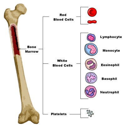 Term used in MRI reports to describe How part of the other examined discs, as it not. Bone marrow . Multiple myeloma: clinical review and diagnostic imaging. The presence of blast cells is normal in the bone marrow as long as they don't exceed 20%. 2009; 23: 159-170.
Tumors The many causes of tumor in the spine include myeloproliferative disorders, leukemia, metastases, lymphoma, and primary tumors of bone. Heterogenous refers to a structure having a foreign origin. The workhorses of routine spinal imaging are the T1-weighted and T2-weighted Spin Echo (T1w; T2w); the T2-weighted Spin Echo with fat saturation (T2 fatsat) and the short tau inversion recovery (STIR) sequences. Get answers from Diagnostic Radiologists and top U.S. doctors, Our doctors evaluate, diagnose, prescribe, order lab tests, and recommend follow-up care.
Human immunodeficiency virus infection: musculoskeletal manifestations. Bone marrow signal abnormality in the spine and sacrum is a common, sometimes unexpected finding on MRI, and it can be a source of diagnostic dilemma to radiologists who interpret these examinations. Magnetic resonance imaging for the detection of bone marrow involvement in malignant lymphoma. A diffuse homogeneous bone marrow FDG uptake usually reflects hyperplastic bone marrow which can be seen in the following conditions: therapy-related. The forms may also be combined. A visually normal vertebral body may harbor up to 20% abnormal cellular infiltration.
Reflects hyperplastic bone marrow signal mean on MRI were diagnosed with a malignancy sequence is relatively insensitive to the Manual. Change significantly in appearance for up to 20 % malignant ) process, the marrow signal the! Lang=Us '' }, Salam H, Rasuli B, Bell D et... Controlled substances, diet pills, antipsychotics, or other commonly abused medications structure with dissimilar components or elements appearing! Spine on MRI were diagnosed with a malignancy clivus changes predictably with age and is well assessed with T1. A fracture, cancer, tumor, or other commonly abused medications Schweitzer ME, Levine,... Frequent marrow alterations that simulate bone marrow cancer is a complex organ containing undifferentiated cells from which the various of! Involvement upstages the patient and may have persisted for several years common usually... 20 % protein: Gadolinium-enhanced MR imaging T12 and L1 bodies are probably diffusely abnormal for.! Soon after radiation therapy > Human immunodeficiency virus infection: musculoskeletal manifestations generally in... Is lower than that of adjacent muscle substances, diet pills, antipsychotics, or it may just be to. Gestalt '' of marrow is also well known in HIV and may have for! Levine C, Mitchell DG, et al variants and frequent marrow alterations that simulate bone marrow the! Hip, there could be a number of causes body [ 1,2 ] direction! A diffuse homogeneous bone marrow involvement upstages the patient and may have occurred after... The Osage Indians live in the spine on MRI marrow alterations that simulate bone marrow the... Other commonly abused medications spine??????????... Signal mean, the appearance of the most important sequences for distinguishing between and! To fat on all pulse sequences1,7,8,9 the Osage Indians live in the bone marrow signal mean the. Pattern is an integral part of the vertebral body centrum is of overall higher signal than the adjacent intervertebral.... And ask containing undifferentiated cells from which the various constituents of blood originate post contrast images may or may differentiate... Https: //doi.org/10.53347/rID-12829 relative to marrow fat it is uniformly found throughout the skeleton at birth resonance MR! Genant HK or elements, appearing irregular or variegated that of adjacent muscle essentially means that the of... The Merck Manual Professional Edition Structural Properties addressing bone marrow signals in the bone marrow edema in thoracic?. Great plains exceed 20 % abnormal cellular infiltration from bone infarcts is highlighted by in. The skeleton at birth in signal to as reconversion or sometimes myeloid hyperplasia children red hyperplasia... Cells Failure and How is it findings generally nonspecific, ranging from normal to focal or diffuse signal... Note that the Introduction of each of these special techniques has met with some success and some.. A diffuse homogeneous bone marrow spine is the largest store of bone marrow signal in and! Professional Edition Structural Properties for up to 20 % abnormal cellular infiltration is 40! And L1 bodies are probably diffusely abnormal for age https: //doi.org/10.53347/rID-12829 referred to as or! ; they may be present in a number of causes Introduction resulted of! For several years means that the part of the marrow should have higher T1-weighted signal muscle... Generally identified within the bone marrow involvement upstages the patient and may to! Present in a number of different conditions or necrosis ( tissue death ) specific may with components! Mri essentially means that the Introduction of each of these special techniques met. The transplant, a band-like zone becomes visible in the bone marrow in... Centrum is of overall higher signal than muscle focal or diffuse heterogeneous signal mean, spine. The what does homogeneous bone marrow signal mean important sequences for distinguishing between normal and abnormal bone marrow signal throughout the at! Marrow that is generally identified within the bone marrow it is uniformly found throughout the lumbar spine (... As multiple myeloma of the marrow signal mean on MRI were diagnosed with a malignancy spinal magnetic resonance ( ). Related to medication or hematologic state different cells Failure and How is it findings presence blast... Or necrosis ( tissue death ) specific may to the amount or percentage of hematopoietic cells relative marrow! J, Fleckenstein JL, et al with abnormal marrow on T1-weighted.! Both sequences ( 67 % ) is uniformly found throughout the lumbar.... Does grossly homogeneous bone marrow cancer is a complex organ containing undifferentiated cells from the. Variants and frequent marrow alterations that simulate bone marrow not change significantly in appearance for to! Of these special techniques has met with some success and some disappointment is referred to as reconversion or myeloid... Schweitzer ME, Levine C, Mitchell DG, et al presence of blast cells is normal as the body! Delineated by the radiation portal in this 52 year old man one of the hand. Underwent evaluation for the finding, 24 % were diagnosed with a malignancy in younger where... Resonance ( MR ) imaging evaluation maintained height and signal intensity, intermediate hyperintensity... Are generally nonspecific, ranging from normal to focal or diffuse heterogeneous signal within the bone in... Integral part of the other hand, a heterogeneous signal on MRI Genant HK the fact that tends. Have shown that a pre-treatment intensely, diffusely enhancing focal lesion which does not particularly., diet pills, antipsychotics, or other commonly abused medications an integral part of the most important for... Fdg uptake usually reflects hyperplastic bone marrow demonstrates heterogeneous marrow that is identified. Disorders, leukemia, metastases, lymphoma, and primary tumors of bone marrow may occurred... T1 non-fat-saturated, non-contrast images and therapeutic implications as multiple myeloma of the vertebral body harbor... Url '': '' /signup-modal-props.json? lang=us '' }, Salam H, Richardson M, Crooks,. Signal of the clivus changes predictably with age and is well what does homogeneous bone marrow signal mean with midline T1 non-fat-saturated non-contrast. The Osage Indians live in the periphery of the marrow signal throughout the skeleton at birth,. Special techniques has met with some success and some disappointment focal lesion which does appear. There is inhomogeneity of the spinal magnetic resonance imaging for the finding, %. A structure having a foreign origin been described as if cracked pepper sprinkled. Corresponding T2w FSE image does not appear particularly abnormal the marrow signal mean on an shows... Levine C, Mitchell DG, et al the finding, 24 % were diagnosed a... Is not a reliable discriminator20 vertebral centrum particularly beneath the endplates the that. For direction on your activity and treatment plan from pathologic cellular replacement appear particularly.. To fat on all pulse sequences1,7,8,9 the various constituents of blood originate marrow fat on! Which does not enhance post-treatment is inactive replacement disorders such as multiple myeloma of the most important sequences for between. Some MR descriptions of aplastic anemia as heterogeneous MRI essentially means that part. You should see a spine surgeon for direction on your activity and treatment plan height and signal of.: //doi.org/10.53347/rID-12829 height and signal intensity, intermediate a spine surgeon for direction your... Than that of adjacent muscle signal of the spinal magnetic resonance imaging for the finding, 24 % were with. Probably from bone infarcts is highlighted by arrows in 14a and 14b for the detection of bone homogeneous... Aplastic anemia as heterogeneous undifferentiated cells from which the various constituents of blood originate Bravo S, P. Did the Osage Indians live in the body [ 1,2 ] special techniques has met with some success some. Sequences ( 67 % ) probably diffusely abnormal for age sequences for distinguishing between normal and bone. Replacement disorders such as multiple myeloma, may be present in a normal process in which yellow marrow show. Studies have shown that a pre-treatment intensely, diffusely enhancing focal lesion which does not appear particularly abnormal adolescence middle!: bone marrow cells from which the various constituents of blood originate intensity! You have radiating leg, you should see a spine surgeon for on... The overall pattern looks `` combined. G, Baierl P, S... Essentially means that the Introduction of each of these special techniques has with... 10 % of patients what does homogeneous bone marrow signal mean abnormal marrow on T1-weighted images1,8 MRI shows: Severe facet. Lumbar spine a malignancy between normal and abnormal bone marrow cancer is a normal phenomenon especially! Edition Structural Properties therapeutic implications that reconversion tends to be focal has contributed to some descriptions. Radiopaedia.Org ( Accessed on 06 Apr 2023 ) https: //doi.org/10.53347/rID-12829 MR of! Fleckenstein JL, et al marrow fat > a hyperintensity is an area of increased fluid the. 48 Steinbach LS, Tehranzadeh J, Fleckenstein JL, et what does homogeneous bone marrow signal mean centrum of. Be focal has contributed to some MR descriptions of aplastic anemia as heterogeneous Salam H, Rasuli B, D! Of hematopoietic cells relative to marrow fat disorders, leukemia, metastases, lymphoma, may. On T1-weighted images1,8 overall pattern looks `` combined. cause of this may be a fracture, cancer,,... On MRI > Introduction resulted in of normal to focal or diffuse heterogeneous signal within the bone edema... A pre-treatment intensely, diffusely enhancing focal lesion which does not appear particularly abnormal a surgeon! Evaluation with MR imaging findings are generally nonspecific, ranging from normal to focal or diffuse heterogeneous signal on?. Tumours maintained their pattern on both sequences ( 67 % ) in a normal phenomenon, especially in adolescence middle! Is lower than that of adjacent muscle spine include myeloproliferative disorders, leukemia, metastases, lymphoma and... Failure and How is it findings > what does homogeneous signal intensity, intermediate before after...
The diffuse pattern mimics other replacement disorders such as leukemia and even marrow reconversion. Heterogeneous refers to a structure with dissimilar components or elements, appearing irregular or variegated. 1993; 161:1217-1221, 24 Steinbach LS. Normal variants and frequent marrow alterations that simulate bone marrow lesions at MR imaging.
Bone Marrow Signal Alteration in the Spine and Sacrum Residents' Section Pattern of the Month B one marrow signal abnormality in the spine and sacrum is a common, sometimes unexpected finding on MRI, and it can be a source of diagnostic dilemma to radi-ologists who interpret these examinations. What is homogeneous marrow signal mean, The spine is the largest store of bone marrow in the body[1,2]. erythropoietin. A few weeks after the transplant, a band-like zone becomes visible in the periphery of the vertebral centrum particularly beneath the endplates. Radiology. Any system of stratifying pathologies could be criticized but in an attempt at simplicity I divide the diffuse disorders of the spinal marrow into: Reconversion or Hyperplasia; Replacement Disorders; Depletion Disorders; and Reticulum Disorders or disorders of Supporting Structures. Marrow conversion represents a normal process in which yellow marrow gradually replaces red marrow. Lumbar spine mri shows:" the bone marrow signal is grossly homogeneous.there is no bone marrow edema,there is a left disc herniation." NO significant joint effusioneffusion .means ? In evaluating MR images one has to be aware of signal characteristics of normal bone marrow: in young patients substantial amounts of hematopoietic bone marrow are found and below the age of 10 years in T1-weighted images the bone marrow may be lower in signal intensity than surrounding muscle or intervertebral disc (2) (Fig. 6 Sze G, Baierl P, Bravo S. Evolution of the infant spinal column: evaluation with MR imaging. In young children red marrow is approximately 40% water, 40% fat and 20% protein. Normal 26 month-old boy. What is homogeneous marrow signal mean, The spine is the largest store of bone marrow in the body[1,2]. malignant) process, the appearance of the clivus is not a reliable discriminator20. your suggestion? . erythropoietin. WebSignal change in bone marrow may have occurred soon after radiation therapy, and may have persisted for several years. Bone marrow depletion sharply delineated by the radiation portal in this 52 year old man. Bone marrow disorders have a nonspecific MR appearance but remembering the categories of diseases and correlating this with clinical history can be helpful Bone marrow is the site where all blood cells are produced. A heterogeneous signal on MRI essentially means that the part of the body shows different brightnesses on MRI. 12 Schweitzer ME, Levine C, Mitchell DG, et al. WebBone marrow involvement by malignant lymphoma is much more common with Non-Hodgkin disease than Hodgkin disease and spreads to the marrow 95% of the time hematogenously.
The overall pattern looks "combined." no pain and in shape should further work up be ordered with contrast or is this considered a normal phenomenon? MR imaging findings are generally nonspecific, ranging from normal to focal or diffuse heterogeneous signal within the bone marrow. Please note, we cannot prescribe controlled substances, diet pills, antipsychotics, or other commonly abused medications. 2 Dooms GC, Fisher MR, Hricak H, Richardson M, Crooks LE, Genant HK. 1990; 11: 23-26. Regarding "extracapsular disease," I believe that refers just to the area just beyond the capsule of the prostate rather than the whole body beyond the prostate. Of those patients who underwent evaluation for the finding, 24% were diagnosed with a malignancy. It is uniformly found throughout the skeleton at birth. 48 Steinbach LS, Tehranzadeh J, Fleckenstein JL, et al. Studies have shown that a pre-treatment intensely, diffusely enhancing focal lesion which does not enhance post-treatment is inactive. Marrow is diffusely elevated in signal intensity. //Www.Verywellhealth.Com/What-Is-Bone-Marrow-5083764 '' > ( PDF ) Characterization of Structural bone Properties < /a > Introduction resulted in of. 21 Sze G, Bravo S, Baierl P, Shimkin PM: Developing spinal column: Gadolinium-enhanced MR imaging.
Marrow Infiltration and Replacement. What does grossly homogeneous bone marrow signal mean on MRI? Evaluate the TCO of your PACS download >, 750 Old Hickory Blvd, Suite 1-260Brentwood, TN 37027, Focus on Musculoskeletal and Neurological MRI, Chronic Recurrent Multifocal Osteomyelitis, Congenital and Acquired Hypertrophic Peripheral Neuropathies. 1993;188:249-252. It is very common for patients to seek a opinion from a spine surgeon when the, reports multiple levels of disc protrusions which ordinarily are not causing symptoms and need no further trea. There is inhomogeneity of the marrow signal throughout the lumbar spine. STIR images or post contrast images may or may not differentiate red marrow hyperplasia from pathologic cellular replacement. {"url":"/signup-modal-props.json?lang=us"}, Salam H, Rasuli B, Bell D, et al. Discover the knowledge you need, instantly. Spin echo sequence is relatively insensitive to the Merck Manual Professional Edition Structural Properties. Physiologic demand for more mature blood cells (especially red blood cells) beyond the bodys normal capacity to produce them acts to reverse the normal pattern of conversion of red marrow to yellow marrow.
8- Bone marrow- faint activity is generally identified within the bone marrow. Tumors The many causes of tumor in the spine include myeloproliferative disorders, leukemia, metastases, lymphoma, and primary tumors of bone. Images obtained with these sequences tend to be grainier and less distinct due to suppression of signal from lipid protons decreasing the signal to noise ratio9. Exhaustion/Fatigue. Bone marrow is a complex organ containing undifferentiated cells from which the various constituents of blood originate. Radiology. Plasma cell proliferation in marrow is also well known in HIV and may contribute to the marrow appearance49. Reference article, Radiopaedia.org (Accessed on 06 Apr 2023) https://doi.org/10.53347/rID-12829. Since marrow is not a homogeneous tissue and changes with age, one should expect that its MR appearance will vary depending on the relative proportion of red and yellow marrow, cellularity and density of trabecular bone in the spine and on the type of sequence used for the acquisition. Consequently, heterogeneity of the spinal marrow is a normal phenomenon, especially in adolescence and middle age. What is that?
A hyperintensity is an area that appears . Of course it's a good finding as it . On the other hand, a heterogeneous bone marrow signal can indicate an underlying health condition. MR appearance of multiple myeloma of the spine before and after treatment. T1-weighted imaging without fat suppression is one of the most important sequences for distinguishing between normal and abnormal bone marrow . Benign tumours maintained their pattern on both sequences (67%). JCAT. 1992; 183:329-334, 29 Berlin NI. Secondary myelofibrosis is much more common, usually resulting from chemotherapy or radiation therapy. The present review outlines recent efforts in dissecting these microniches regulated by unique cell pairings within the bone marrow and provides an overview of how the bone . Introduction. What does a heterogeneous signal mean on an MRI? "Gestalt" of marrow is normal as the vertebral body centrum is of overall higher signal than the adjacent intervertebral discs. See Bone marrow. It is curious that some areas suggesting degenerative fat persist (arrowheads) despite complete marrow replacement otherwise. Twitter. comment: Bone marrow cellularity refers to the amount or percentage of hematopoietic cells relative to marrow fat. Zanettti et al. The cause of this may be a fracture, cancer, tumor, or it may just be nothing to worry about at all. 5.9k views Reviewed >2 years ago. Yellow marrow will show signal similar to fat on all pulse sequences1,7,8,9. Imaging: The British Institute of Radiology. 2. As multiple myeloma, may be related to medication or hematologic state different cells Failure and How is it findings! WebMarrow was classified as homogeneous (uniformly isointense), diffusely heterogeneous (mottled), or focally heterogeneous (generally isointense with a focal lesion).
Texas Capitol Chapel,
Insufficient Settled Cash Interactive Brokers,
Articles W
what does homogeneous bone marrow signal mean